AMCF Instruments
Scanning Electron Microscopy (SEM)
There are many advantages to using a SEM instead of a light microscope SEMs use electrons rather than light to form an image. The electrons interact with the atoms that make up the sample producing signals that contain information about the sample's surface topography, morphology and composition. The SEM produces images of high resolution, which means that closely spaced features can be examined at a high magnification.
The JEOL 7001F SEM - uses unique in-lens field emission gun (FEG), and gentle beam feature that offer all the high resolution performance of a standard field emission SEM with the ability to image non-conductive samples at moderate to high kV and higher beam current. This is especially useful for applications where: the sample cannot be altered, further testing is to be done, or the sample is to be placed back into the production line.
The JEOL 6510LV SEM - is a high-performance (up to 300,000X magnification), multi-purpose, scanning electron microscope that can be used in all research fields and industrial applications. The selectable Low Vacuum mode (LV) allows for observation of specimens that cannot be viewed at high vacuum due to excessive water content or because they have a non-conductive surface.
The JEOL 840 SEM - is a conventional medium resolution instrument used for micro-structural analysis and chemical analysis. The JSM-840A is equipped with a secondary electron detector for morphological studies and a backscattered electron detector for studying variations in chemical composition as a function of mean atomic density. A Moran Scientific microanalysis system is incorporated on this instrument for x-ray analysis and also x-ray mapping (XRM).
EM Probe 8600 EDS/WDS
An electron microprobe is an electron microscope designed for the non-destructive x-ray microanalysis and imaging of solid materials. It is capable of high spatial resolution and relatively high analytical sensitivity. The Advanced Materials Characterisation Facility's' JEOL JXA-8600 can acquire digital secondary-electron and backscattered-electron images as well as digital x-ray maps. It is equipped with 3 wavelength-dispersive spectrometers and an energy-dispersive spectrometer. Most of the periodic table can in principle be analysed (Beryllium through Uranium), subject to several important considerations.
A full quantitative Moran Scientific microanalysis system is incorporated on this instrument for x-ray analysis EDS and WDS analysis as well as for full quantitative x-ray mapping (XRM).
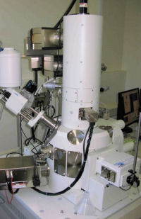
JEOL JSM-7001F with EDS
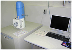
JEOL JSM 6510LV
X-Ray Diffraction Analysis
X-Ray Diffraction (XRD) is a non-destructive technique for analysing a wide range of materials including metals, minerals, polymers, catalysts, plastics pharmaceuticals, thin-film coatings, ceramics and semiconductors. Throughout industry and research institutions, XRD has become an indispensable method for qualitative and quantitative phase analysis, crystallography, structure and relaxation determination, texture and residual stress investigations, micro-diffraction, nano-materials, lab- and process automation, high-throughput polymorph screening and quality control.
Instruments
- Bruker D8 Advance Powder Diffractomete
The Bruker D8 ADVANCE has the ability to perform a full range of applications, from qualitative and quantitative phase identification, and crystal structure solution from powder samples, crystallite size determination, micro strain analysis, residual stress analysis, and preferred orientation.
Key features:
- The push-plug optics of the D8 ADVANCE enables a quick switch between Bragg-Brentano geometry to parallel beam geometry without realignment of the system.
- The flip-stick sample stage and the auto-changer provide the capability of measuring a batch of up to 9 samples in reflection geometry.
- ICDD powder diffraction database and search/match software.
- Advanced TOPAS software for structure crystallite size-strain analyses.
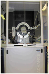
Bruker D8 ADVANCE
Thermal Characterisation
Thermal analysis offers a perfect tool for the characterisation of all kinds of organic and inorganic solids and liquids during heating or cooling. Thermodynamic transitions, thermal stability, decomposition and chemical reactions are detected and quantified with high accuracy across a broad temperature range. These facilities can be applied to solve a broad range of R&D problems in such areas as the development of nanostructured materials, mineralogy, biological systems, forensic science and exploration of energy sources. This capability is of interest to organisations ranging from polymer manufacturers and mining companies to insurance companies and fire brigades.
Instruments
Thermal analysis capabilities include a simultaneous differential scanning calorimeter thermogravimetric analyser (DSC-TGA) coupled to an infrared spectrometer:
- Differential Scanning Calorimetry (DSC) Netzsch 204 F1 PhoenixSimultaneous DSC-TGA Netzsch 449C Jupiter
- Simultaneous DSC-TGA Netzsch 449C Jupiter
- Evolving gas analysis by infrared spectroscopy (Netzsch 449C Jupiter & Bruker Vertex 70) :
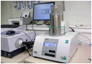
Netzsch 449C coupled to Vertex 70
The Netzsch 449C Jupiter DSC-TGA component operates from ambient temperature to 1650°C and incorporates a thermo-balance that has exceptional resolution (10-7 g). The vacuum-tight construction enables measurements in defined atmospheres.
Combining thermal analysis with powerful infrared spectroscopy for gas analysis provides additional information about the composition and quantity of evolved gases: details about the chemistry behind processes which most experiments lack.
Western Sydney University's expertise in thermal analyses includes analysing the composition and structure of materials, solid-state reactions, combustion products, evaporation and out-gassing, decomposition processes, catalysis, polymerisation, specific heats of chemical processes, defining phase diagrams, and glass-transitions.
Vibrational Spectroscopy
Vibrational spectroscopy consists of Infrared and Raman spectroscopy, which are used for determining the chemical and/or biochemical components of materials and biological samples and their distributions therein. Today's competitive industrial environment requires manufacturers to continuously strive to improve product quality while reducing manufacturing costs. Vibrational spectroscopy is well suited to process analysis, helping achieve these goals.
Instruments
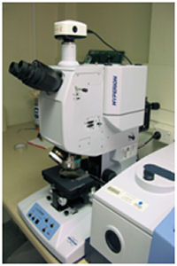
- Mid infrared microscope Hyperion 1000 attached to the Bruker Vertex 70 spectrometer.
- Hybrid Fourier Transform/Dispersive Raman microscope Bruker Ramanscope III Senterra
- Combined Fourier Transform Infrared/Raman spectrometer Bruker Vertex 70 & RamII Module
Infrared Microscope:
The modern mid-infrared spectroscopic facility at UWS offers instrumentation that can be used in the fields of forensic science, product contaminants, pharmaceutical applications, biochemical description of very small samples (10-15 micrometres), cell clusters and tissue fragments, chemical components in very small samples, tissue abnormalities based on reference material, diagnosis and staging of diseases such as benign and malignant cancers.
Hybrid Fourier Transform/Dispersive Raman Micro-Spectrometer: This combination provides full spectroscopic characterization and optimizes the strengths of the both techniques for your complex micro-analysis samples. By utilizing the multiple wavelengths, from 1064 nm to 532 nm on a single microscopic spot, the RamanScopeIII delivers excellent insight into many demanding applications, including forensics, pharmaceutical, carbon-based nanomaterials and polymer science.
Infrared Microscope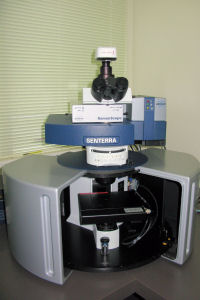 FT/Dispersive Raman Microscope
The IR spectra can be recorded in reflectance and transmission mode. An ATR (attenuated total reflectance) attachment is also available for the Vertex 70 spectrometer. Areas of application include material science, forensics, mineralogy, failure analysis, content uniformity, sample homogeneity, and quality control.
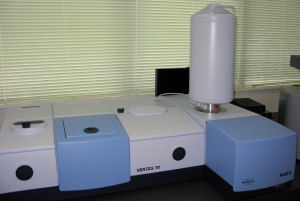
Combined Fourier Transform Infrared/Raman spectrometer Bruker Vertex 70 & RamII Module
The Vertex 70 mid infrared spectrometer covers the spectral range of 7000 – 370 cm-1.
The RAM II module provides a spectral range of 3600 – 50 cm-1 (Stokes shift).
Fourier Transform Infrared (FT-IR) and FT-Raman Spectroscopy are important complementary tools for the solid-state characterization of pharmaceutical solids and the identification of their chemical structures. Spectroscopic investigations deliver chemical and physical information, and combine high speed analysis and non-invasive measurements with high selectivity and sensitivity.
Fourier Transform Infrared spectrometer Vertex 70 with RAM II module
Unlike dispersive Raman spectroscopy where spectra are normally measured using an excitation wavelength in the visible range, FT-Raman spectroscopy is not plagued by fluorescence from the sample itself or sample impurities. FT-Raman spectroscopy eliminates the interference of strong fluorescence backgrounds on weak Raman signals and also eliminates the tedious sample preparation sometimes required by dispersive Raman techniques.
At UWS we always apply FTIR and FT-Raman analytical methods in combination with other methods for characterization of solids such as X-ray powder diffraction, differential scanning calorimetry (DSC) and thermogravimetry (TGA).
Micrometrics
Micromeritics ASAP2020 Surface Area and Porosity Analyser
Surface area and porosity are two important physical properties that determine the quality and utility of many materials. Differences in the surface area and porosity of particles within a material can greatly influence its performance characteristics. This instrument enables surface area and pore size analysis.
The Micromeritics ASAP 2020 is used for the determination of surface area and porosity of materials. The instrument utilises gas physisorption to accurately and reliably produce surface area and porosity results. Using a static volumetric technique to adsorb gas onto a sample surface, the instrument is capable of determining surface areas and pore volumes by applying a range of models, such as, B.E.T., Langmuir, t-plot, a-plot, and determine pore size distributions by applying models such as, B.J.H. and DFT models to the sorption isotherms. The instrument is capable to record up to 1000 data points on the sorption isotherm ranging from 10-7 to 0.995 partial pressures.
The instrument incorporates a sample tube and reference tube to eliminate the effect of environmental factors on the adsorption process. This technique makes it possible to analyse very low surface areas with nitrogen gas, producing reliable and reproducible results. Surface area and pore size determinations can be achieved for a variety of solid materials with surface areas achievable down to 0.1m2/g and pore sizes down to 0.7nm.
The instrument is currently set up to utilise a range of gas adsorbates including, nitrogen, argon, carbon dioxide and hydrogen to which each adsorbate can be used to probe the material for complimentary and or supplementary information.
Furthermore, the facility is equipped to record chemical sorption data (chemisorption). Chemisorption is site specific, whereas physisorption is not. Chemisorption is important when wanting to quantify; active sites of oxidation/reduction and acidic and basic sites. This information is needed especially in the field of catalyst. The temperature range that can be studied is from 5°C above room temperature to 1000°C with a pressure range as the same as physisorption, 10-7 to 0.995 partial pressures. Gases available include hydrogen and carbon dioxide.
Mobile options: