You can search for courses, events, people, and anything else.
Two imaging technologies are available in the BMRF, magnetic resonance imaging (MRI) which uses magnetic fields and magnetic field gradients, and microcomputed tomography (MicroCT) imaging which uses X-rays. It is possible to reconstruct 3D images of suitable samples using these imaging technologies. MR imaging can also be performed in several modalities, e.g., diffusion weighted/tensor imaging or mapping of relaxation time constants, to gain more information about the samples. The MicroCT in the BMRF allows for high speed/low dose imaging.
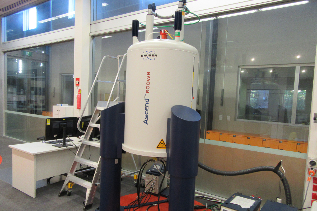
BRUKER AVANCE IIIHD 600 MHz Wide Bore NMR/MRI Spectrometer (14.1 T)
Equipped with high field XYZ gradient amplifiers capable of generating up to 30 T/m (dependent on probe). It can be equipped with various probes and accessories that enable it to be used for a wide range of NMR experiments such as heteronuclear NMR, diffusion, and micro-imaging.
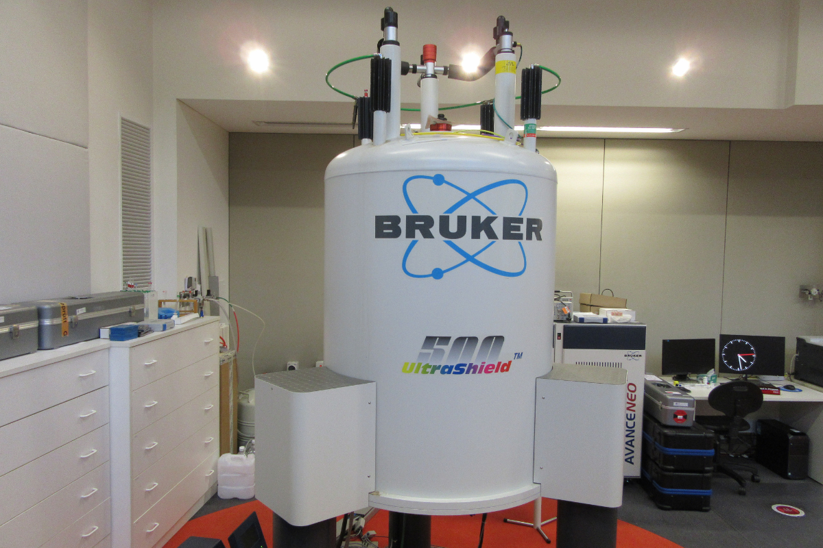
BRUKER AVANCE NEO 500 MHz Wide Bore NMR/MRI Spectrometer (11.7 T)
Equipped with high field XYZ gradient amplifiers capable of generating up to 30 T/m (dependent on probe). It can be equipped with various probes and accessories that enable it to be used for a wide range of NMR experiments such as heteronuclear NMR, diffusion, micro-imaging, and high-resolution MAS.
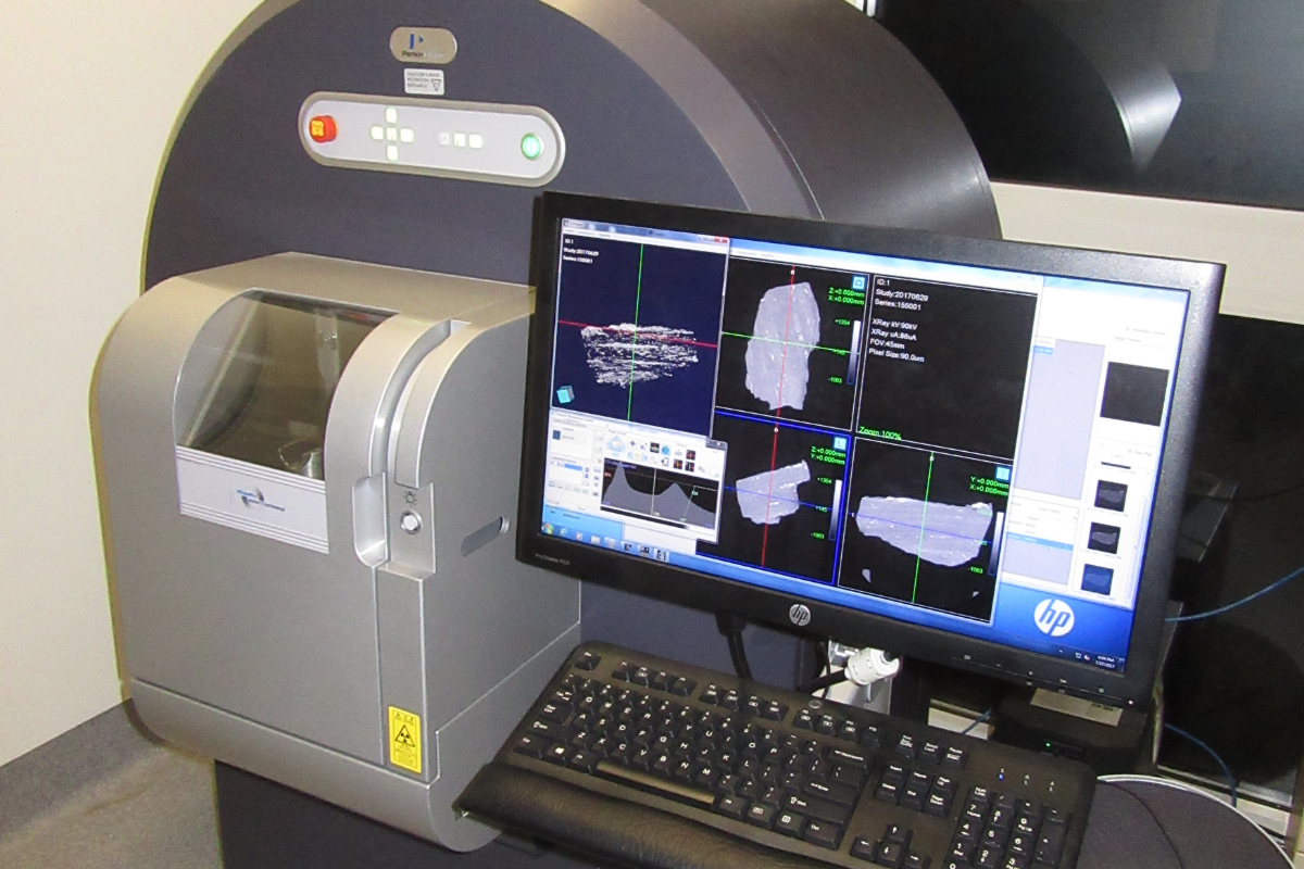
PerkinElmer QUANTUM GX MicroCT
This imaging system provides high-resolution computed tomography images. The Quantum GX can image with a very high resolution (4.5 µm). This scanner can image with a wide field of view (FOV) (e.g., 36 mm, 72 mm).